


Summary:Researchers at Johns Hopkins University have developed an autonomous robotic system that enables precise retinal vein cannulation with AI-driven control, high-resolution imaging, and micrometer-scale accuracy. The technology achieves high success rates in simulated conditions and could advance safer, more reliable treatments for retinal vein occlusion and other delicate eye diseases.
On January 20th, scientists at Johns Hopkins University in the United States developed a new autonomous robotic system that promises to precisely perform retinal vein cannulation, opening new avenues for treating serious eye diseases. This research, based on advanced robotic control technology, integrates high-resolution imaging technology and deep learning algorithms, demonstrating outstanding perception and feedback capabilities. The relevant findings were published in the latest issue of the journal “Science Robotics”.
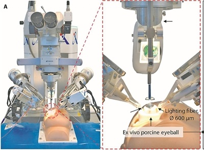
[Image caption: Schematic diagram of the experimental setup. Image source: Phys.org]
Retinal vein occlusion is a blinding eye disease. When veins in the retina—the photosensitive tissue at the back of the eye—are blocked, it leads to a rapid decline or even loss of vision. Currently, retinal vein cannulation is considered a highly promising treatment. Doctors precisely insert an extremely fine needle into the blocked vein and inject thrombolytic or anti-angiogenic drugs. However, the diameter of the retinal vein is roughly the same as the thickness of a human hair, requiring extremely high surgical precision and making manual operation extremely difficult; even the slightest mistake can damage the fragile retina.
A newly developed autonomous robotic system can stably perform cannulation tasks at the micrometer scale, ensuring accurate needle entry into the target blood vessel while minimizing tissue damage. Studies show that the robot achieves a 90% success rate in cannulation within a static eye and maintains an 83% success rate under dynamic simulation conditions. Crucially, the system can reliably identify when the needle tip contacts and successfully enters the vein.
The system is equipped with two robots specifically designed for minimally invasive ophthalmic surgery, one holding the needle and the other the manipulation tool. The hardware integrates three deep learning models, which, after training, can track needle tip movement in real time, identify anatomical structures, and autonomously plan the optimal path to ensure precise puncture. Currently, the team has conducted tests on isolated pig eyes, with some experiments simulating the periodic micro-movements of the eyeball caused by in vivo respiration.
The team states that the maneuvering precision required for retinal vein cannulation is less than 100 micrometers, far exceeding the physiological capabilities of the human hand. This breakthrough demonstrates that, with the help of robotics and artificial intelligence, the most delicate ophthalmic surgeries can now be partially automated, while maintaining safety, accuracy, and reproducibility. However, before it can be truly applied in clinical practice, its effectiveness and safety still need to be verified in live animal and human trials.

